
TCR Sequencing: Decoding the Immune Response Across Vaccines, Cancer Immunotherapy, and Disease
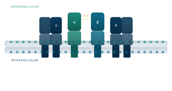
Every T cell in your body carries a unique molecular fingerprint: the T-cell receptor (TCR). This receptor determines which antigens a T cell can recognise and sequencing it at scale means you can read the history and activity of an immune response with precision. Which clones expanded? Which are actively transcribing? Which survived checkpoint inhibitor treatment to attack a tumour?
TCR sequencing has moved from a specialised research curiosity to an essential translational tool. It is now being used to assess vaccine breadth, stratify patients before immunotherapy, track tumour-infiltrating lymphocytes (TILs), and annotate epitope-specific responses, all from a blood draw or tissue biopsy.
This post explores the key applications of TCR sequencing, why the choice of RNA versus DNA input matters, and how single-cell and cell-aggregate approaches are recommended for different biological questions.
The expanding applications of TCR sequencing
Measuring vaccine breadth and depth
An effective vaccine generates a durable, broad T-cell response. But traditional assays (ELISpot and flow cytometry with tetramers) only tell you whether a response exists against a handful of known epitopes. TCR sequencing tells you how many clones responded, how broadly they diversified, and how long they persist.
By comparing the TCR repertoire before and after vaccination, researchers can quantify clonal expansion, assess repertoire diversity, and identify which clonotypes are enriched by vaccination. This is especially critical for next-generation mRNA vaccines, cancer vaccines targeting neoantigens, and therapeutic vaccines for infectious disease, where breadth of response often predicts durability of protection.
Patient stratification in checkpoint inhibitor therapy
Immune checkpoint inhibitors (ICIs), including anti-PD-1, anti-CTLA-4, and related therapies, have transformed oncology. Yet only a subset of patients responds. Identifying responders before treatment, or early during the treatment cycle, remains one of the most pressing challenges in immuno-oncology.
TCR sequencing is a powerful stratification tool. Studies have shown that the T-cell repertoire at baseline, and its dynamics after treatment initiation, differ significantly between responders and non-responders. Key metrics include:
- Clonality: more oligoclonal repertoires at baseline may signal pre-existing immune activation
- Clonal expansion dynamics: responders often show early clonal expansion of tumour-specific T cells after ICI initiation
- Clonal replacement vs. revival: new tumour-infiltrating clones (replacement) versus re-activation of pre-existing ones (revival) are both associated with response, but with different kinetics and mechanisms
Unlike PD-L1 expression or tumour mutational burden (TMB) measurements, both static snapshot biomarkers, TCR dynamics offer a longitudinal window into whether immunotherapy is working.
Monitoring anti-tumour immune responses
In solid tumours, TILs are recognised as prognostic, but not all TILs are equal. TCR sequencing of TILs allows researchers to identify which clonotypes are tumour-reactive, whether they are expanding or exhausted, and how treatment shifts the balance.
When combined with matched blood samples, circulating and tumour-resident TCR clones can be tracked longitudinally. This non-invasive monitoring strategy is becoming central to clinical trial design in immuno-oncology.
RNA or DNA? Sensitivity vs. quantification in TCR sequencing
One of the most underappreciated decisions in TCR sequencing study design is whether to use RNA or genomic DNA (gDNA) as input material. Both provide critical biological insight but are suited for different questions.
DNA-based TCR sequencing: precision cell counting
When you sequence TCR genes from gDNA, you are counting the number of T cells carrying each unique CDR3 sequence. Since each T cell carries one rearranged TCR per haplotype, and the number of DNA copies is proportional to cell number, DNA-based sequencing provides a quantitative, cell-count-proportional view of the repertoire.
Advantages:
- Direct proportionality between clonotype frequency and T-cell abundance
- Detects both expressed and unexpressed TCR alleles, as gDNA contains both alleles even though T cells typically express only one after allelic exclusion
- Stable template: less susceptible to pre-analytical degradation
Limitations:
- Lower sensitivity for rare clonotypes compared to RNA-based methods
RNA-based TCR sequencing: capturing active immune responses
RNA sequencing targets TCR transcripts, and because activated T cells dramatically upregulate TCR transcription, RNA-based approaches capture immunologically active clonotypes.
Advantages:
- Higher sensitivity: each T cell contains multiple RNA copies of its TCR, making RNA methods more effective at detecting low-frequency clonotypes
- Functional relevance: only expressed transcripts are captured, reflecting true biological specificity: what the T cell is actually doing
- The preferred first-line method for FFPE analysis when DV300 thresholds are met
Limitations:
- RNA is less stable than DNA and requires careful pre-analytical handling
Single-cell vs. cell-aggregate TCR sequencing: resolution vs. breadth
The next major methodological axis is the choice between single-cell and cell-aggregate (bulk) sequencing. These are not competing approaches; they are complementary lenses.
Bulk sequencing analyses the entire TCR repertoire of a sample simultaneously, generating millions of CDR3 sequences. It excels at:
- Repertoire diversity analysis: comparing clonal richness and evenness across samples or timepoints
- Tracking clonal dynamics: measuring expansion, contraction, or persistence of specific clones across treatment cycles
- Cost-effective cohort-scale studies: bulk sequencing is orders of magnitude cheaper per sample than single-cell approaches, enabling statistically powered clinical studies
The limitation is that bulk sequencing lacks paired chain information (TCRα and TCRβ together), which is critical for identifying the precise receptor responsible for antigen recognition.
The trade-off is depth: standard single-cell approaches capture thousands to tens of thousands of cells per sample, while bulk methods routinely sequence millions of TCR molecules, enabling detection of many more clonotypes.
Which approach to choose?
| Biological question | Recommended approach |
|---|---|
| How broad is the vaccine response? | Bulk (RNA preferred) |
| Which patients are likely to respond to ICI? | Bulk & longitudinal sampling |
| What is the phenotype of the tumour-reactive clone? | Single-cell |
| What TCR should we clone for adoptive T-cell therapy? | Single-cell |
In many translational programs, the right answer is a phased design: use bulk sequencing for cohort-wide screening and biomarker discovery, then apply single-cell sequencing to samples of interest identified in the bulk phase.
TCR epitope annotation: bridging sequence and antigen recognition
Knowing which T cells are present, how many, and how they change in relation to therapy is powerful, but the final question is: what antigen are they recognising?
TCR epitope annotation links specific CDR3 sequences (or full αβ TCR pairs) to their target antigens using curated databases combined with computational prediction tools. At TATAA, we offer this capability through our partnership with ImmuneWatch, whose DETECT platform and proprietary IMWdb database are purpose-built for this challenge.
IMWdb is the largest curated TCR–epitope database covering more than 200,000 TCRs across over 2,400 unique epitopes, with quarterly updates. Coverage spans oncology antigens (including KRAS mutants, MART1, WT1, NY-ESO-1), infectious diseases (SARS-CoV-2, CMV, EBV, Influenza A, HIV, HBV), and autoimmune conditions (Type 1 Diabetes, Multiple Sclerosis, Celiac Disease).
When epitope annotation is applied on top of TCR sequencing data, the result is transformative: a clonotype expanded in tumour tissue can be identified, linked to a known neoantigen or viral peptide, and assessed for functional relevance, all within a single analytical framework. For vaccine and immunotherapy studies, this allows researchers to determine not just that T cells expanded, but which specific epitopes drove the dominant response, enabling rational immunogen design and therapy monitoring.
What this means for your research program
Whether you are running a Phase I/II clinical trial of a therapeutic cancer vaccine, developing a patient stratification strategy for checkpoint inhibitor therapy, or investigating tumour-infiltrating lymphocyte biology, TCR sequencing gives you a mechanistic, quantitative read on the adaptive immune response. The same capability extends to preclinical settings, including syngeneic tumour models, adoptive cell transfer studies, and vaccine immunogenicity testing in mouse, where early characterisation of T-cell responses can inform and de-risk clinical program design.
A well-designed TCR sequencing study does not just add data to your program; it provides the mechanistic evidence that regulatory agencies, scientific reviewers, and translational partners increasingly expect.
TATAA Biocenter’s TCR sequencing services
TATAA offers comprehensive TCR sequencing services, supporting both DNA and RNA input, bulk and, through our partner, single-cell workflows. Our qualified workflows, from extraction to downstream bioinformatic analysis, are designed for clinical and preclinical applications, including GCLP-adherent studies across patient samples.
We use Cellecta’s DriverMap platform for high-sensitivity CDR3 profiling, with detection capability extending to rare, low-frequency clonotypes from limited input material. Where study design requires additional flexibility, we can implement other targeted TCR amplification kits, for example the Takara SMART-Seq TCR kit, to accommodate specific sample types, input amounts, or continuity with your existing datasets.
Repertoire data is analysed using MiXCR, a gold-standard bioinformatics platform for immune repertoire profiling from NGS data. It enables accurate V(D)J alignment, robust CDR3 extraction, and clonotype assembly from raw reads, supporting quantitative analysis of diversity and clonal dynamics. Its optimised, preset-driven pipelines and broad compatibility across different datasets make it a robust standard for reproducible immune monitoring.
Talk to our immune monitoring team to discuss the right TCR sequencing design for your program.
What is TCR sequencing used for?
TCR sequencing is used to analyse the diversity, clonality, and dynamics of T-cell receptor repertoires. Applications include monitoring vaccine-induced immune responses, stratifying cancer patients for immunotherapy, tracking tumour-infiltrating lymphocytes, and identifying antigen-specific T-cell clones.
Should I use RNA or DNA for TCR sequencing?
DNA-based TCR sequencing is proportional to T-cell number and stable, but detects both expressed and unexpressed alleles. RNA-based TCR sequencing is more sensitive, captures only actively expressed receptors, and better reflects functional immune activity. Running both simultaneously provides the most complete picture.
What is the difference between bulk and single-cell TCR sequencing?
Bulk TCR sequencing profiles the entire repertoire at once, enabling deep clonotype detection and cost-effective cohort studies. Single-cell TCR sequencing provides paired TCRα/β chain information alongside gene expression data per cell, enabling phenotype-linked clonotype analysis. Both approaches are complementary.
What is TCR epitope annotation?
TCR epitope annotation is the process of linking CDR3 sequences to their target antigens, using databases of known TCR–peptide–MHC interactions and computational similarity methods. It is used to determine the antigen specificity of expanded T-cell clones.
Can I couple TCR repertoire analysis to immune response-related proteomic markers?
Yes. Integrating TCR sequencing with protein-level immune profiling adds an important functional dimension to repertoire data, linking clonal dynamics to the cytokine and immune mediator environment in which T cells are operating. This multi-modal approach is particularly powerful for biomarker discovery in immuno-oncology and for stratifying patients in clinical trials.
We support this through affinity-based proteomics using the Olink platform, including the Inflammation and Immune Response panels, covering cytokines, chemokines, and checkpoint-related markers. For the most comprehensive view, the Illumina Protein Prep (IPP) profiles 9,300+ targets, enabling discovery-driven analysis alongside targeted immune monitoring.
